Oleksandr Kosenko
surgeon-proctologist of the highest category:when your back end is in safe hands
chief physician of the medical center «LEFT BANK PROCTOLOGY CENTER»
- Conservative and surgical treatment of hemorrhoids, anal fissure, pilonidal sinus (pilonidal cyst), rectal fistula, rectal hernia, rupture, etc.
- Minimally invasive removal of a polyp, cyst, or tumor.
- High effectiveness.
- Treatment of adults and children.
- “Left Bank Proctology Center”: when your back end is in safe hands!
Kyiv, Livoberezhna metro station, Rusanivka residential area, 49 Entuziastiv Street.
Experience: over 25 years of work in the field of proctology.
Doctor’s phone: +380 50 330 15 15
Doctor’s Opinion
Hello! I am a surgeon-proctologist, just as specific a person as the rest of surgeons. The term “proctologist” is associated by many people with the word hemorrhoids. Hemorrhoids are the most common reason for visiting a proctologist. Recently, doctors have achieved success in this field – removal of hemorrhoids by bio-welding or excision of a rectal fissure with a laser has become a new generation operation and is displacing outdated methods.
But few people know that bleeding from the anal canal can be not only a symptom of hemorrhoids or fissure, but also more dangerous problems, including such as a polyp or rectal tumor. A surgeon-proctologist helps to figure out these issues. The list of primary examinations performed by a proctologist includes digital examination, rectoscopy (rectoromanoscopy), fibrocolonoscopy, biopsy if necessary – all this is called endoscopy.
But let’s not talk about alarming things, we’ll discuss that separately. Today, the patient is concerned with two main questions – the quality and price of surgery to remove hemorrhoids or fissure. The outcome of the disease often depends on the surgery method. The patient is increasingly concerned about the question – will a monopolar or bipolar coagulator, radio wave knife, ultrasonic or laser scalpel, bio-welding clamp be used? At the same time, few people are interested in the qualification of the surgeon-proctologist, work experience in surgery.
When searching on the Internet, people usually ask: good proctologist, best surgeon-proctologist, Kyiv proctologists rating and reviews, proctology for women, recommend a good proctologist, proctologist consultation 24/7. Sometimes, succumbing to advertising tricks, the patient goes on a long path of ineffective treatment or uses dubious technologies. Proctological problems should be solved by a specialist with the appropriate category and work experience.
I draw attention to the fact that people often search for “Proctology Center of Ukraine”, “Ukrainian Proctology Center”, “Proctological Center of Ukraine” in queries. The correct philologically-based name is “Ukrainian Coloproctology Center”. The center is located at the proctology department of Kyiv City Clinical Hospital No. 18, but I no longer work there. Now I work as a surgeon-proctologist and chief physician at a medical center called “LEFT BANK PROCTOLOGY CENTER”, located at: Kyiv, 49 Entuziastiv Street, near Livoberezhna metro station (picturesque Rusanivka district). To book an appointment, call: +380 50 330 15 15
- Attention: we work even without power supply, the operating room is powered by our own inverter batteries.
- Download the rules for preparation for proctologist examination.
How Are Proctological Diseases Treated?
Conservative Therapy
Medication Treatment in Proctology
Medication treatment is the foundation of conservative therapy for many proctological diseases and often becomes the first step to recovery. Properly selected drug therapy can significantly alleviate symptoms of hemorrhoids, anal fissures, paraproctitis and other pathologies, and in some cases completely eliminate the problem.
Local medications form the basis of symptomatic treatment for proctological diseases. Suppositories, ointments and gels act directly at the site of inflammation, providing quick relief from pain, itching, and discomfort. Suppositories are convenient to use and effective for internal hemorrhoids or rectal inflammation, as the active substance is evenly distributed across the mucosa. Ointments and gels are better suited for treating external hemorrhoids, anal fissures, and perianal dermatitis.
Modern local medications contain a combination of active substances: anti-inflammatory components reduce swelling and redness, local anesthetics relieve pain, venotonics improve blood circulation in hemorrhoidal veins, and healing agents accelerate tissue regeneration. It is important to understand that the effect of local treatment appears quickly, but a prolonged course is needed to consolidate the result.
Systemic medications affect the body as a whole and are especially useful for chronic forms of diseases. Phlebotonic agents strengthen vein walls, improve venous outflow, and reduce congestion in the pelvic organs. These drugs are particularly effective for hemorrhoids, as they eliminate the very cause of hemorrhoidal node enlargement. Anti-inflammatory drugs help control the inflammatory process, while analgesics ensure patient comfort during treatment.
A separate group consists of laxatives and stool regulators, which play a key role in the prevention and treatment of many proctological problems. Hard fecal masses traumatize the rectal and anal canal mucosa, provoking fissure formation and hemorrhoid exacerbation. Mild laxatives based on lactulose or macrogol not only facilitate defecation but also improve gut microflora.
It should be emphasized that medication treatment is most effective at early stages of the disease, in combination with dietary recommendations and lifestyle changes. When selecting medications, individual patient characteristics, concomitant diseases, and possible contraindications are always taken into account, so avoid self-treatment – consult a proctologist.
Diet Therapy in Proctology
Proper nutrition is an integral part of treatment and prevention of proctological diseases. Diet therapy can significantly alleviate symptoms, accelerate recovery, and prevent relapses of many rectal and anal canal pathologies.
Dietary fiber plays a key role in maintaining healthy bowel function. Soluble fibers found in vegetables, fruits, oatmeal, and legumes form a gel-like mass that gently stimulates peristalsis and creates optimal fecal consistency. Insoluble fibers from whole grains, nuts, and seeds increase the volume of intestinal contents and accelerate their movement through the colon.
Special attention should be paid to prebiotics – substances that nourish beneficial gut microflora. Inulin found in chicory, Jerusalem artichoke, and onions, oligofructose in bananas and asparagus, pectins in apples and carrots create favorable conditions for bifido- and lactobacteria growth. Healthy microflora not only improves digestion but also strengthens local immunity, reducing the risk of inflammatory processes.
Drinking regimen is no less important for preventing proctological problems. Sufficient fluid intake – at least 30 ml per kilogram of body weight per day – ensures normal fecal consistency and prevents constipation. Pure water, non-acidic compotes, and weak herbal teas are especially beneficial. It is important to distribute fluid intake evenly throughout the day rather than drinking large amounts at once.
Dietary restrictions aim to eliminate irritants and reduce inflammation. Spicy food, alcohol, and strong coffee stimulate blood circulation in the pelvis and can trigger hemorrhoids or anal fissures exacerbations. Fatty, fried foods slow peristalsis and contribute to constipation. Sweets, especially those with refined sugar, disrupt gut microflora balance.
Instead of prohibited foods, it is better to prefer easily digestible food: boiled, stewed, or baked dishes, fermented dairy products, lean meats, and fish. Meals should be regular, in small portions, with thorough chewing.
It should be emphasized that the diet must be balanced and maximally varied. Radical restrictions can harm overall health, so all dietary changes should be coordinated with a proctologist, especially if there are concomitant digestive system diseases.
Physiotherapy in Proctology
Hardware treatment methods have become an integral part of modern proctological practice, especially when medications do not produce the desired result. Physiotherapeutic effects restore functions of the affected area without surgery, making it an attractive option for many patients.
Electrical stimulation of pelvic floor muscles has proven effective for recovery after various proctological problems. Weak electrical discharges activate weakened muscle fibers, restoring their normal contraction ability. This method is particularly valuable for women after complicated childbirth, elderly patients with bowel incontinence, and those who have undergone perineal surgery. Typically, 9-15 procedures are needed to achieve noticeable improvement.
Ultrasound therapy affects tissues through high-frequency mechanical waves that penetrate deep into perineal structures. This treatment activates blood supply to venous plexuses, relieves muscle tension, and accelerates damaged tissue recovery. Patients with hemorrhoidal nodes report reduced pain and swelling, while with anal canal fissures healing occurs faster than usual.
Magnetic field therapy in treating proctological patients is based on its ability to improve blood circulation in small vessels. Special equipment creates controlled magnetic fields of varying intensity that favorably affect cellular metabolic processes and reduce inflammation. The procedure is painless and suitable even for patients with increased sensitivity to other physiotherapy types.
Low-intensity laser irradiation is used for targeted effects on problematic mucosal and skin areas. Laser beam of specific wavelength stimulates intracellular recovery processes, accelerates collagen and elastin synthesis needed for normal healing. In case of anal defects, laser can completely eliminate pain and prevent coarse scar formation.
A course of physiotherapy procedures usually lasts from one to two weeks, depending on the specific situation and the body’s response to therapy. Tolerance to such treatment is high, serious adverse reactions are rare. However, there are application limitations: active tumor processes, acute infections, heart rhythm disorders, presence of electronic implants in the body.
Experience shows that hardware physiotherapy is particularly useful in complex cases when conventional treatment does not produce the expected effect. Individual parameter selection and combination of different methods often yield striking results, allowing patients to return to full life.
Blockades in Proctology
Therapeutic blockades are an effective method of pain relief and anti-inflammatory treatment in proctology, allowing quick elimination of pain syndrome and improvement of patients’ quality of life. This method is particularly valuable for chronic pain syndromes, anal sphincter spasm, and postoperative period.
Novocaine blockades are used for temporary pain relief and diagnostic purposes. Novocaine blocks nerve impulse conduction, providing complete anesthesia of the injection area. For anal fissures, novocaine blockade is performed pararectally, eliminating sphincter pain spasm and creating conditions for mucosal defect healing. The procedure is outpatient, effect occurs within minutes and lasts 2-4 hours. Treatment course usually consists of 5-7 injections with 2-3 day intervals.
Corticosteroid blockades combine pain-relieving effects of local anesthetics with powerful anti-inflammatory action of hormonal drugs. Diprospan, kenalog, or hydrocortisone are most commonly used in combination with lidocaine or novocaine. Such blockades are particularly effective for perineal inflammatory processes, chronic coccyx pain, postoperative scar changes. Steroids reduce tissue swelling, suppress inflammatory reaction, and improve tissue nutrition.
It is important to understand that corticosteroid blockades have longer action – from several weeks to months, but cannot be performed too frequently due to tissue atrophy risk. Usually 1-3 injections with 2-4 week intervals are sufficient.
Sacral (caudal) blockades are performed in the sacral canal area and intended for pain relief of the entire perineum and pelvic organs. This method is used for complex pain syndromes when pain spreads throughout the pelvic area, genital nerve neuralgia, chronic pelvic pain. Sacral blockade is technically more complex than other types, requiring precise anatomical knowledge and doctor’s experience.
The procedure is performed under palpation or ultrasound control for accurate needle positioning. A mixture of long-acting local anesthetic (marcaine, naropin) and corticosteroid is used. Sacral blockade effect can last from several months to a year, making this method particularly valuable for chronic pain syndromes.
All types of blockades have contraindications: allergic reactions to anesthetics, infectious processes at injection site, blood clotting disorders, severe systemic diseases. After the procedure, the patient is monitored for 30-60 minutes to control possible complications.
It should be emphasized: blockades are not only symptomatic treatment but also a therapeutic method that can break the “vicious circle” of pain sensations and spasm. Properly performed blockade often becomes a turning point in treating chronic proctological diseases and allows avoiding more complex surgical interventions.
Minimally Invasive Treatment Methods in Proctology
Mechanical Methods of Hemorrhoid Treatment
Mechanical approaches in hemorrhoid therapy are based on blocking blood flow to affected nodes, causing their contraction and gradual elimination. Such interventions belong to the category of minimally traumatic procedures performed in clinic conditions.
The most commonly used is elastic latex ring ligation. The essence of the manipulation lies in placing rubber rings around the base of pathologically altered nodes using a special instrument under anoscope control. The rubber ring compresses the arteries supplying the hemorrhoidal node, as a result of which it loses nutrition and is rejected independently within a week to ten days. This approach is justified for the second-third stage of internal hemorrhoids when formations can still be reduced into the anal canal.
An alternative is the technique of surgical thread ligation. The principle of action remains unchanged, but using threads instead of latex allows finer regulation of the compression degree and suits people with rubber intolerance. The manipulation is performed under local anesthesia within a quarter of an hour.
Main indications for mechanical methods of hemorrhoid treatment include cases of internal hemorrhoids with bleeding episodes when nodes prolapse but return to place independently or after manual intervention. Among limitations of mechanical methods are acute thrombosis of nodes, presence of anal ruptures, inflammatory processes in the rectal area, and blood clotting system problems.
In the post-procedure period, patients often complain of moderate unpleasant sensations and the feeling of a foreign object in the anal area for several days. Patients are recommended a balanced diet with sufficient plant fibers, restriction of heavy physical work for the first five days, and careful maintenance of cleanliness in the problem area. With proper patient selection, effectiveness of mechanical methods reaches eighty-ninety percent.
Chemical Methods of Hemorrhoid Treatment
Chemical methods of hemorrhoid treatment are based on using special preparations that cause sclerosis (shrinkage) of hemorrhoidal nodes or their coagulation. These methods are particularly effective at early stages of the disease and with small node sizes.
Sclerotherapy involves injecting a sclerosing agent directly into the hemorrhoidal node or submucosal base around it. The most commonly used are solutions of ethanolamine oleate, polidocanol, or sodium tetradecyl sulfate. These substances cause aseptic inflammation of vessel walls, leading to their gluing and obliteration. The procedure is performed under anoscope control, the drug is injected with a thin needle in the amount of 1-3 ml depending on node size.
Chemical coagulation involves using more aggressive chemical agents that cause tissue necrosis of hemorrhoidal node. Such preparations include phenol in oil solutions, trichloroacetic acid, or polyinol. This method requires special caution due to the risk of burns to surrounding tissues and is used less frequently than sclerotherapy.
Advantages of chemical methods include simplicity of execution, no need for special equipment, minimal pain, and quick recovery. The patient can return to normal activities the next day. These methods are most effective for internal hemorrhoids of stage 1-2 with bleeding but without significant node prolapse.
Contraindications to chemical methods include allergy to sclerosing agents, acute inflammatory processes in the anorectal area, thrombosis of hemorrhoidal nodes, and pregnancy. After the procedure, short-term pain, burning sensation, and minor spotting may occur for several days.
Sclerotherapy effectiveness is 60-80% depending on disease stage and technique correctness. In some cases, a repeat procedure may be needed after 4-6 weeks to achieve optimal results.
Physical Energy Impact Methods
Physical energy impact methods in proctology use various types of energy for coagulation (coagulation) of hemorrhoidal node tissues. These methods provide precise control of impact depth and minimal damage to surrounding healthy tissues.
Infrared photocoagulation is based on converting infrared spectrum light energy into thermal energy. A special device generates infrared radiation with a wavelength of 1200 nm focused on hemorrhoidal node tissue. Under heat action, protein coagulation and vessel obliteration occur, supplying the node. The procedure is performed through anoscope, exposure duration is 1-2 seconds per point.
Laser coagulation uses a focused laser beam of various wavelengths (most commonly diode laser 980 nm or neodymium laser 1064 nm). Laser radiation causes instant tissue coagulation with scab formation that falls off in 7-10 days. This method allows very precise energy dosing and penetration depth control.
Bipolar electrocoagulation applies high-frequency electric current for tissue heating and coagulation. Unlike monopolar coagulation, the bipolar method is safer as current flows only between two electrodes of the instrument, not spreading throughout the patient’s body.
Radio wave coagulation (Surgitron-type devices) uses high-frequency radio waves of 3.8-4.0 MHz. This method provides the softest tissue impact with minimal thermal damage and fastest healing.
All energy impact methods are performed under local anesthesia, last 10-15 minutes, and allow patients to immediately return to normal activities. They are most effective for internal hemorrhoids stage 1-2. After the procedure, minor discomfort for 2-3 days and superficial scab formation that falls off independently are possible. Effectiveness of these methods is 70-85%, and complication risk is minimal with proper technique execution.
Latest Hemorrhoid Treatment Methods
Modern proctology actively implements innovative hemorrhoid treatment methods that combine high effectiveness with minimal trauma. These technologies represent a new approach to treatment aimed not only at symptom elimination but also at restoring anatomy and function of the anorectal area.
HAL-RAR (Hemorrhoidal Artery Ligation with Rectoanal Repair) is a revolutionary method using Doppler ultrasound for precise localization of arteries supplying hemorrhoidal nodes. A special proctoscope with an ultrasound sensor allows the surgeon to detect all arterial branches and ligate them under sound signal control. After stopping blood supply, mucopexy is performed – pulling up and fixing prolapsed mucosal tissues to the rectal wall with special sutures.
Advantages of HAL-RAR include no tissue removal, minimal postoperative pain, and quick recovery. The method is effective for all stages of internal hemorrhoids, including the fourth stage with constant node prolapse. The procedure is performed under local or spinal anesthesia, lasts 30-40 minutes, and the patient can be discharged the same day.
PRP-therapy (Platelet-Rich Plasma Therapy) is based on using the patient’s own blood plasma enriched with platelets and growth factors. Plasma is obtained by centrifugation of patient’s venous blood, then injected directly into hemorrhoidal nodes or surrounding tissues. Platelets release biologically active substances that stimulate tissue regeneration, improve microcirculation, and reduce inflammation.
PRP-therapy is particularly effective at early stages of hemorrhoids and can be used as an independent treatment method or in combination with other procedures. The method is absolutely safe since it uses the patient’s own cells, eliminating allergic reactions or infection transmission. Treatment course usually consists of 2-3 procedures with 2-3 week intervals.
Latest methods require special equipment and proctologist training, but provide better long-term results with minimal recurrence risk. They are particularly indicated for patients with concomitant diseases requiring gentle surgical intervention and young people for whom quick return to active lifestyle is important.
An individual approach to treatment method selection is recommended depending on disease stage, patient age, concomitant pathology, and personal preferences. Modern minimally invasive technologies allow effective hemorrhoid treatment with minimal discomfort and quick return to normal life.
Surgical Treatment
Classical Surgical Operations in Proctology
Classical surgical interventions in proctology remain the foundation of treatment for many rectal and anal canal diseases, despite the development of modern technologies. These operations have stood the test of time and demonstrate high effectiveness with proper indication selection and technique execution.
Hemorrhoidectomy is the radical removal of hemorrhoidal nodes used for the third and fourth stages of the disease. Milligan-Morgan operation involves complete excision of hemorrhoidal nodes leaving open wounds that heal independently. Ferguson method differs by suturing wounds after node removal, which accelerates healing and reduces postoperative pain syndrome. Both techniques ensure reliable long-term results but require qualified execution and proper postoperative rehabilitation.
Hemorrhoidopexy, or Longo operation, is an alternative method for hemorrhoid treatment, particularly for rectal mucosa prolapse. The essence of the operation consists of removing a circular strip of mucosa above the hemorrhoidal nodes while simultaneously pulling tissues back into the anal canal. This method is significantly less painful compared to classical hemorrhoidectomy and allows patients to return to normal life faster.
Sphincterotomy is used to treat anal fissures, especially chronic ones. The operation involves cutting part of the internal anal sphincter, which reduces pressure in the anal canal and creates conditions for fissure healing. The intervention can be performed either openly or closed, depending on the clinical situation.
Fistulectomy is an operation to remove anal fistulas (fistulae) that can complicate the course of paraproctitis. The surgical technique depends on the fistula tract location relative to the sphincter and can range from simple opening of superficial fistulas to complex reconstructive interventions for high transsphincteric fistulas.
It should be emphasized that the choice of classical surgical method is always individual and depends on the disease nature, process stage, concomitant pathology, and patient’s general condition. Modern anesthesiology allows these interventions with minimal discomfort, while proper postoperative rehabilitation ensures quick recovery and return to full life.
Modern Surgical Technologies in Proctology
Medical innovations of recent decades have fundamentally transformed the treatment of rectal and anal canal diseases. New techniques significantly reduce surgical trauma, shorten recovery period, and alleviate postoperative pain compared to standard surgical procedures.
BIWELD tissue electrofusion technology opens fundamentally new possibilities for joining biological structures. The method works through high-frequency electrical impulses that create molecular bonds between tissues, reducing the need for suturing threads. In proctological practice, this approach ensures reliable hermetic tissue connection, reduces bleeding and infection probability, while significantly accelerating postoperative recovery.
Radio wave surgical systems like Surgitron or “FOTEK” operate based on high-frequency radio signals for tissue cutting and cauterization. The main advantages of this technique lie in exceptional cutting precision, preservation of surrounding healthy areas, absence of electric current in patient’s body, and accelerated healing of surgical wounds. Radio wave interventions demonstrate the best results for removing condylomas, papillomas, and small formations around the anus.
Ultrasonic surgical instruments use sound vibration power for tissue dissection with simultaneous hemostasis. This technology ensures exceptional intervention precision along with effective bleeding control, which is critically important during operations on highly vascular intestinal wall tissues. Ultrasonic instrumentation allows surgeons to work even in the narrowest anatomical spaces, avoiding thermal damage to adjacent structures.
Argon plasma coagulation is classified as a non-contact tissue cauterization method using electrically charged argon gas. The method proves extremely useful for stopping mucosal bleeding, treating hemorrhagic intestinal lesions, and destroying superficial pathological formations. The technology allows controlling impact depth and virtually eliminates the risk of intestinal wall perforation.
The mentioned techniques are used both separately and in combination with traditional surgical interventions, creating opportunities for personalized treatment of each patient. It should be remembered that the choice of specific technology is determined by disease characteristics, pathological focus location, and surgeon-proctologist‘s professional skills in working with appropriate equipment.
Reconstructive Surgery in Proctology
Reconstructive surgery in proctology occupies a special place as it aims to restore anatomy and function of the anal canal and rectum after trauma, inflammatory processes, oncological diseases, congenital anomalies, and previous operations. These interventions require high surgeon qualification and deep understanding of pelvic floor anatomy.
Anoplasty is an operation to restore the anal canal used for scar strictures, congenital anomalies, or after traumatic injuries. The essence of the intervention lies in reconstructing the anal opening using local tissues or grafts. The operation may include cutting scar tissues, using skin-mucosal flaps, and forming a new anal canal with restoration of its elasticity and functionality.
Sphincteroplasty is performed to restore anal sphincter integrity after childbirth trauma, iatrogenic injuries during previous operations, or trauma. The operation involves using ends of damaged muscle followed by their connection using special sutures. Intervention success largely depends on the time elapsed since trauma, degree of scar changes, and general condition of sphincter muscle tissue.
Promontofixation is a complex operation for treating complete rectal prolapse, especially in elderly patients. The intervention can be performed either via open access or laparoscopically. The essence of the operation consists of freeing the rectum, pulling it up, and fixing it to the sacral promontory using synthetic mesh. This restores normal pelvic floor anatomy and eliminates prolapse.
Reconstructive operations are often combined with pelvic floor plasty, especially in women with combined pelvic organ prolapses. In such cases, a comprehensive approach with simultaneous correction of several anatomical structures anatomy may be used. The cosmetic effect of reconstructive operations (cosmetic proctology) is also important.
A feature of reconstructive surgery in proctology is the need for prolonged rehabilitation and stepwise function restoration. Patients are often prescribed special therapeutic exercises to strengthen pelvic floor muscles, biofeedback therapy for sphincter coordination recovery, and pharmacological support.
Success of reconstructive operations depends not only on technical intervention execution but also on proper preoperative preparation, including functional examination of pelvic floor muscles, and comprehensive postoperative rehabilitation under multidisciplinary specialist team control.
Laparoscopic and Endoscopic Interventions in Proctology
Minimally invasive surgical techniques are today recognized as the best treatment option for numerous rectal diseases, combining excellent therapeutic results with minimal body harm. Laparoscopic and endoscopic operations enable even the most complex interventions through small incisions or natural body openings, dramatically reducing hospital stay and recovery time.
Laparoscopic resections have found wide application for rectal cancer, diverticulosis, complete rectal prolapse, and certain types of intestinal inflammatory processes. The surgical process occurs through 4-5 small openings in the abdominal wall through which a video camera and special instruments are inserted. The laparoscopic method provides unparalleled visualization of internal structures, enables the most precise tissue manipulation with minimal blood loss, and preserves nerve fibers controlling pelvic organ function.
Transanal endoscopic microsurgeries TEM and minimally traumatic interventions TAMIS represent the most modern developments for removing benign and early-stage malignant intestinal formations. These approaches enable removal of neoplasms located up to 15-20 cm from the anal ring, avoiding major traumatic operations with partial bowel resection. The procedure is performed through the natural anal passage using specialized endoscopic equipment that ensures maximum tumor removal precision with minimal risk of adverse consequences.
TEM technique is based on using a special operating rectoscope with optical system and working channels that enables performing the most delicate manipulations on the intestinal wall under constant visual control. TAMIS represents an advanced version of this technique using standard laparoscopic instrumentation and disposable port, making the technique more accessible for routine use.
Endoscopic submucosal dissection ESD is used for removing large superficial neoplasms of the rectum and lower sigmoid colon segments. This technique allows complete tumor removal in one piece, ensuring the most accurate morphological diagnosis and reducing disease recurrence possibility, unlike partial removal.
Laparoscopic operations for diverticular pathology involve removing the diseased bowel segment followed by connecting healthy sections, which can be performed electively for recurrent diverticulitis or emergently for complications. The minimally invasive surgical approach is particularly beneficial for young patients and recurrent pathology forms.
Combined laparoscopic-endoscopic interventions create the possibility of simultaneous bowel mobilization laparoscopically and neoplasm removal endoscopically, significantly expanding organ-preserving operation capabilities.
Effectiveness of minimally invasive interventions substantially depends on proper patient selection, quality preoperative examination, and operating team professionalism. Such technologies require specialized equipment and long-term training but provide patients with higher quality of life and quick return to full activity.
Postoperative Support and Rehabilitation
Medical Support
The postoperative period after proctological surgery requires close attention from medical staff. Each patient goes through an individual recovery path that depends on intervention complexity and body characteristics. Experience shows that properly organized medical supervision significantly shortens recovery time.
Pain management begins already on the operating table and continues throughout the entire healing period. The anal area has a dense network of sensitive nerves, so discomfort can be pronounced. Combination of different groups of pain medications gives the best result. Local anesthetics block pain impulses directly in tissues, anti-inflammatory agents reduce swelling, and for severe pain they add opioid analgesics. Interestingly, effective pain relief not only alleviates suffering but actually accelerates tissue recovery by preventing muscle spasm.
Complication prevention requires constant vigilance from the medical team. The wound in the anal area is prone to infection due to proximity to the intestine with its microflora. Antibacterial therapy is prescribed taking into account the risk of each specific case. The surgeon conducts thorough thrombosis risk assessment, especially in elderly patients and those with heart and vascular problems. Wound care includes daily dressings with special solutions and monitoring for alarming symptoms.
Systematic examinations allow tracking recovery dynamics and timely intervention if needed. The first visit usually occurs one to two days after intervention, subsequent ones – after a week, a month, and three months. During each meeting, the proctologist checks wound condition, adjusts prescriptions, and consults on regimen issues. Sometimes special optical devices are used for detailed result examination, allowing assessment of mucosa healing quality. Such systematic approach prevents problems at early stages.
Physical Rehabilitation
Return to normal motor regime after proctological intervention occurs gradually. The body needs time to restore not only tissues but also functional connections between different systems. Properly structured physical rehabilitation program becomes the foundation for complete recovery.
Special motor exercises begin already in the first days after surgery. Breathing exercises activate blood circulation in the small pelvis and prevent congestion. Gradually, perineal and abdominal muscle exercises are added. Special place is occupied by pelvic floor strengthening techniques, including Kegel method. These exercises teach the patient to consciously control sphincter work and coordinate their activity. The instructor selects load individually, taking into account patient’s condition and surgery type.
Hardware treatment methods significantly accelerate recovery process. Warm sitz baths with medicinal solutions soothe tissues and reduce inflammation. Laser irradiation stimulates cell regeneration, magnetic fields improve local blood circulation. Electrophoresis delivers medications directly to affected area. Ultrasound softens scar tissue and restores muscle fiber elasticity. Each procedure has its indications and contraindications, so appointment is made after thorough examination by proctologist.
Sphincter apparatus functional recovery often becomes the most difficult rehabilitation stage. These muscles are responsible for holding intestinal contents and controlled defecation. Training includes tension and relaxation exercises for different muscle groups with gradually increasing duration and contraction strength. Biofeedback allows patient to see results of their efforts on monitor screen. Electrical stimulation helps “remind” muscles of proper work rhythm. The whole process may take several months, but persistence is usually rewarded with complete function restoration.
Psychological Support
Patient’s emotional state after proctological surgeries affects recovery speed no less than surgical intervention quality. Many people are embarrassed to talk about sensations in this delicate area, creating additional internal tension. Experienced proctologists always dedicate time to talk with patient about what they feel and experience.
Qualified psychologist assistance is sometimes simply irreplaceable. Some patients experience real horror before first defecation after surgery, imagining terrible pain. Others worry about how surgery will affect their personal life and whether disease will return. Specialist teaches simple relaxation techniques, helps replace negative thoughts with positive ones, and sets up for quick recovery. Best results are given by methods combining conversation with practical exercises to overcome fears.
Recovery period after surgery is often accompanied by feeling of vulnerability and anxiety. For many patients it’s important to feel they are not left alone with their experiences. Warm attitude of medical staff, family support, and opportunity to share fears help overcome psychological barrier. Simple realization that anxiety and embarrassment are natural feelings reduces tension level. Open conversation about emotions, stress management advice, and in some cases light group or individual consultations create atmosphere of trust. This psychological atmosphere gives patient confidence that path to complete recovery will be calmer and significantly shorter.
Treatment result depends on how well patient and doctor understand each other. Strict adherence to all recommendations, timely visits to proctologist, and belief in success – this is what is needed for effective and complete recovery. Modern medicine gives us powerful tools for treating proctological diseases, but patient’s own desire to regain health remains particularly important factor.
What Does Proctologist Oleksandr Kosenko Treat?
Hemorrhoids
Hemorrhoids are one of the most common proctological diseases that occur due to varicose expansion of rectal veins. In a proctologist’s practice, hemorrhoids are encountered daily: most patients seek help already at the stage of complications – for example, with node thrombosis or prolapse.
hemorrhoids can be internal, external, or combined. Internal nodes are hidden inside the canal, external ones are under the skin near the anus, both forms can combine and give complex symptoms. For internal hemorrhoids, an international classification (stages I-IV) is accepted – from slight enlargement to node prolapse that does not reduce independently.
Main factors of hemorrhoid development:
-
chronic constipation;
-
sedentary lifestyle;
-
excessive physical exertion;
-
pregnancy and childbirth;
-
abuse of spicy food or alcohol;
-
prolonged sitting.
Under the influence of these factors, pressure in pelvic vessels increases and inflammation develops.
Hemorrhoid symptoms to which special attention should be paid:
-
appearance of blood during or after defecation (bright red, without mucus or stool admixture);
-
itching, sensation of foreign body, burning in anal area;
-
acute pain, especially with node thrombosis;
-
gradual enlargement/prolapse of node during straining.
Among complications there are massive bleeding with anemia development, node incarceration with necrosis, secondary infection, as well as formation of anal skin tags that complicate hygiene.
Hemorrhoid diagnosis includes examination by proctologist and instrumental studies (anoscope, rectoscopy) – this not only determines hemorrhoid stage but also excludes other pathologies, including polyps and tumors. Important to remember: not every bleeding is hemorrhoids. Self-diagnosis in this case is dangerous.
Hemorrhoid treatment depends on disease severity. At early stages lifestyle changes are needed (fiber-rich diet, sufficient water, physical activity, hygiene), local medications and venotonics. For more serious forms minimally invasive methods (latex ligation, infrared coagulation) or classical surgery are used.
Special attention is needed for pregnant patients – gentle treatment methods are recommended for them, with emphasis on diet and hygiene.
Hemorrhoids do not pose direct threat to life, but significantly affect its quality and can mask more dangerous diseases. Therefore prevention, timely examination, and risk factor control are the key to your health.
Anal Fissure
Anal fissure is a longitudinal defect of anal canal mucosa that occurs due to tissue rupture or damage. Such rupture leads to formation of painful wound, often accompanied by sphincter spasm and acute pain during defecation. Most often fissure is located on posterior wall of anal canal, less often – on anterior or lateral walls.
Causes of anal fissure are varied, but main ones are: chronic or acute constipation, mucosa traumatization during passage of hard fecal masses, enhanced spasm of internal anal sphincter, inflammatory processes in intestine, as well as various infections, intestinal and venereal diseases.
Clinical picture of anal fissure includes:
-
acute pain during and after defecation that can last from several minutes to several hours;
-
bright red blood discharge on toilet paper or in stool;
-
itching, burning in anus area;
-
feeling of anxiety and discomfort due to pain and inability of complete defecation;
-
in chronic cases induration (scar tissue) may appear near fissure, sphincter spasm becomes more pronounced.
Diagnosis of anal fissure consists of rectal palpation examination, anoscopy and rectoromanoscopy to clarify damage localization and depth, as well as exclusion of concomitant pathologies (hemorrhoids, tumors, proctitis). Colonoscopy is prescribed if needed to exclude colon diseases.
Anal fissure treatment depends on disease duration and course character.
Acute anal fissure usually responds well to conservative therapy that includes:
-
diet high in fiber (fruits, vegetables, bran) and sufficient fluid intake to soften stools;
-
constipation elimination using mild laxatives;
-
use of local remedies – antispasmodic and analgesic ointments and suppositories (for example, nitroglycerin ointment, “Relief”, “Posterisan”);
-
sitz baths with chamomile decoction or antiseptic solutions to reduce inflammation and pain;
-
mild microclysters to relieve pain and accelerate healing.
Chronic anal fissure (over 3 months) often requires surgical treatment by proctologist, since scar tissue forms in tissues making conservative therapy ineffective. Most common surgeries are:
-
lateral internal sphincterotomy – small incision of internal sphincter to relieve spasm and improve blood circulation in fissure area;
-
fissurectomy – excision of affected mucosa area together with scar tissues;
-
modern minimally invasive methods – laser or radio wave fissure removal that are painless and have short rehabilitation period.
Rehabilitation after anal fissure removal surgery requires diet adherence, prevention of physical exertion and regular proctologist control to avoid recurrences.
Prevention of anal fissure consists of ensuring regular soft stools, avoiding mucosa traumatization during defecation, constipation and diarrhea control, treatment of inflammatory bowel diseases.
Pilonidal Cyst
Epithelial coccygeal tract (pilonidal cyst) is a condition manifested by formation of a small cavity or canal in the coccyx area, under the skin between the buttocks. Inside, hair, skin particles, and sebum often accumulate, creating conditions for inflammation. Some patients discover the problem accidentally – during a doctor’s visit or routine medical examination, but much more often the pathology announces itself with pain, redness, and swelling in the coccyx area, and sometimes with pus discharge from small skin openings.
Causes of pilonidal cyst can be varied. Most typically it is associated with hair ingrowth under the skin, constant friction or microtraumas during prolonged sitting, especially if work or lifestyle is sedentary. Anatomical features also contribute – deep intergluteal fold, increased sweating, congenital tissue structure.
Inflammation can develop gradually or acutely. Initially, the patient notices only mild discomfort, but later the skin reddens, swelling and pain appear, especially during sitting or movement. If infection joins, body temperature rises and abscess forms. In chronic cases, fistulas form from which pus or blood periodically discharges, and the process can last for years.
Pilonidal cyst diagnosis is usually established by proctologist during examination. Additional examination methods such as soft tissue ultrasound or fistulography are prescribed when it is necessary to determine the depth and direction of the tract or presence of multiple communicating cavities.
Treatment of pilonidal cyst depends on disease stage. If cyst doesn’t bother and doesn’t inflame, observation and preventive measures may suffice, but complete problem elimination is possible only surgically. Surgery involves removal of epithelial tract together with all affected tissues. Modern surgery offers both classical methods with excision and wound healing, and minimally invasive technologies – laser or endoscopic intervention that give faster results and less postoperative pain. In case of acute suppuration, primary task is abscess opening and drainage, and tract removal is performed after inflammation reduction.
Pilonidal cyst prevention consists of thorough coccyx area hygiene, avoiding prolonged sitting without breaks, sweating control, and if possible – reducing hair coverage in risk area. Timely visit to proctologist at first signs of inflammation allows avoiding more complex surgeries and prolonged rehabilitation.
Polyp
Polyps of the rectum – these are small growths on the mucosa that can be completely benign or turn out to be a pre‑cancerous condition. Most often they appear as soft formations on a stalk or a broad base, which protrude into the bowel lumen. Some of them cause no symptoms for many years and are detected incidentally during examination. Others, in contrast, become noticeable through bleeding, mucus, or discomfort in the anal canal area.
The main danger lies in the fact that some polyps, especially adenomatous ones, have the potential to gradually transform into malignant tumor. Sometimes a villous polyp that has caused no complaints for years begins to grow rapidly and change its cellular structure. This is why physicians recommend not postponing examination, even if symptoms are minimal.
From my own practice I can say: patients often come “just for a check‑up” because of slight blood admixture in the stool, and a polyp requiring removal is found. There are many such cases, and they demonstrate that early diagnosis saves from much more complex treatment in the future.
The most reliable detection method – is colonoscopy. It allows even very small formations to be seen and removed immediately. Modern colonoscopy is a sufficiently safe procedure, and its benefit is enormous. After polyp removal, the tissue is sent for histology – to determine the type of formation and its potential for malignant transformation.
Treatment of a polyp consists mainly in – it must be removed. In most cases this is done endoscopically. However, if there are many formations or they are large, it may be necessary to operate on a larger bowel segment.
Thus, although polyps are not always an immediate danger, they are always a signal for action. In my center, there have been several patients who, after polyp removal, practically avoided an oncological diagnosis later. Remember that preventive examination by a proctologist and an endoscopist after the age of 40 (and earlier if there is a family history) is the best way to avoid colorectal cancer and protect your bowel health.
Tumors of the Rectum and Colon: Benign and Malignant
In daily proctological practice, we often deal with intestinal neoplasms, most of which cause no symptoms to the patient in early stages. From a medical perspective, a tumor is pathological tissue overgrowth resulting from disrupted cell division control. The main thing every patient should know: tumors can be benign or malignant, with significantly different prognosis and risks.
Benign tumors include polyps, lipomas, fibromas, hamartomas. Intestinal polyps are the most common. Their main danger lies in the fact that certain polyp types carry risk of malignant transformation – primarily adenomatous and villous ones. Benign tumors typically:
-
have clear boundaries;
-
do not invade adjacent tissues;
-
do not metastasize;
-
can remain asymptomatic for a long time.
However, even a small polyp can cause hidden intestinal bleeding, anemia, or discomfort if located in distal (far) sections and gets traumatized.
Malignant tumors (rectal or colon cancer) is a serious oncological pathology ranking among the leading causes of cancer mortality worldwide. Cancer is characterized by aggressive growth, invasion of adjacent tissues, ability to metastasize to liver, lungs, lymph nodes.
Main symptoms of malignant intestinal tumor:
-
prolonged or recurrent rectal bleeding;
-
change in bowel movement character (constipation, diarrhea, mucus or blood admixture);
-
feeling of incomplete defecation, tenesmus – false defecation urges;
-
pain, especially at late stages;
-
general exhaustion, weight loss, weakness.
Unfortunately, early stages of malignant tumors are often asymptomatic, therefore regular preventive proctologist examinations are crucial – especially after 45-50 years or with family predisposition.
Diagnosis of malignant intestinal tumors is based on comprehensive approach: clinical history analysis, examination by proctologist, endoscopic studies (rectoromanoscopy, colonoscopy), tumor biopsy, as well as imaging methods (ultrasound, MRI, CT) to assess disease spread.
Colon cancer treatment strategy depends on tumor nature and stage. Benign polyps are usually removed endoscopically with subsequent histology. If malignancy is confirmed, surgical treatment is prescribed, often combined with chemo- and/or radiation therapy. Prognosis depends on treatment method choice, timeliness of seeking help, and patient individual characteristics.
Therefore, be attentive to appearance of blood and even minor changes in bowel movements – these symptoms require diagnosis. In my many years of practice, I have repeatedly seen how early tumor diagnosis enabled organ-preserving treatment and prevented development of severe complications, preserving quality and length of life. That’s why I urge all patients to undergo regular preventive examinations, instrumental screening methods, and consultation with proctologist at the slightest complaints.
Rectal Prolapse
Rectal prolapse (rectal prolapse) is a condition in which part or entire segment of rectum everts and protrudes beyond anal opening. This pathology can develop at any age, but is more common in older patients and women after childbirth.
Causes of rectal prolapse: combination of pelvic floor weakness, chronic constipation, increased intra-abdominal pressure, pelvic trauma, neurological diseases, as well as hereditary predisposition. Significant role is also played by frequent childbirths with trauma, bowel motility disorders, and straining during defecation.
Main symptoms of rectal prolapse include sensation of foreign body or fullness in anal canal, pain in anal area, mucus or blood discharge, as well as clearly expressed tissue prolapse during or after defecation. This pathology is often accompanied by loss of defecation control – fecal or gas incontinence, which significantly worsens patients’ quality of life.
Sometimes prolapse occurs only during straining, but in severe cases – even at rest. If prolonged, complications are possible such as inflammation and swelling of prolapsed tissues, erosions, necrosis, as well as development of chronic pain syndrome.
Diagnosis of rectal prolapse includes examination by proctologist with digital examination, anoscopy or rectoscopy. To clarify size, degree of damage, and assess functional state, additional methods are used: anorectal manometry, anal electromyography, pelvic floor ultrasound, and if indicated, X-ray.
Treatment of rectal prolapse depends on prolapse stage and severity. At early stages conservative management is possible, including stool normalization through high-fiber diet, sufficient fluid intake, mild laxatives, as well as physiotherapy and special exercises to strengthen pelvic floor muscles. Manual reduction of prolapsed tissues with hygiene rules helps avoid infectious complications.
However, it should be understood that conservative therapy for rectal prolapse is usually temporary and often doesn’t solve the problem permanently. In most cases with disease progression, surgical intervention is indicated. Surgeries can vary: from fixation of distal (far from digestive tract beginning) rectum segment to reconstructive techniques with bowel pulling up and pelvic floor strengthening.
Modern proctological techniques allow effectively eliminating rectal prolapse with minimal risk, as late treatment leads to complications including necrosis, constant inflammation, and secondary infection.
I emphasize: if rectal prolapse symptoms are present, it’s important to consult proctologist in timely manner. Proper diagnosis and comprehensive treatment approach significantly improve prognosis and patient’s quality of life.
Perineal Infections, Urethral Infections, Gynecological Infections (Candidiasis, Chlamydia, Herpes, Dermatitis, etc.)
Perineal infections, urethral infections, and gynecological infections are a common problem handled by proctologists, gynecologists, and urologists. These are inflammatory processes that affect the skin and mucous membranes in the perineal area, urethra, and external and internal genital organs. They may be caused by bacteria, viruses, or fungi and are often sexually transmitted or triggered by a microbiome imbalance.
How do infections develop? Disruption of the natural protection of the skin and mucous membranes (due to excessive moisture, microtrauma, hypothermia, or weakened immunity) creates favorable conditions for pathogens to multiply.
Perineal infections may start with mild itching or redness but quickly progress into pronounced inflammation.
Urethral infections (urethritis) often cause acute symptoms in men – burning, pain, discharge – while in women, they may go unnoticed until complications appear.
Gynecological infections – candidiasis, bacterial vaginosis, chlamydia, genital herpes – show diverse courses: from mild yet persistent symptoms to serious reproductive issues.
The most common conditions include:
-
candidiasis (thrush), caused by the overgrowth of Candida fungi, often after antibiotics or hormonal changes, accompanied by curd-like discharge, itching, and swelling;
-
chlamydia – a sneaky infection that often begins without symptoms but over time affects the fallopian tubes, increasing the risk of infertility;
-
genital herpes – a viral disease with periodic painful eruptions that remains in the body for life after infection;
-
dermatitis of the perineum may be allergic or infectious in origin and are often combined with secondary bacterial or fungal involvement;
-
bacterial and viral urethritis – if untreated, may spread to the bladder, prostate (in men), or cervix (in women).
Diagnosis and treatment of perineal, urethral, and gynecological infections must consider that an accurate diagnosis cannot be made based on symptoms alone – the clinical picture of these infections often looks similar, but the causes differ. Therefore, tests such as swabs from the affected area, PCR, bacterial cultures, and sometimes immunological or serological testing are essential. This approach helps identify the pathogen and select targeted therapy – antibacterial, antiviral, antifungal, or combined. Treatment is always accompanied by hygiene recommendations, dietary adjustments, and microbiome restoration.
Prevention of these infections involves not only avoiding random sexual contact and using condoms but also regular check-ups, attention to any changes in the anogenital area, and timely treatment of even minor irritations. With careful self-care, it is possible to prevent chronic forms of gynecological, urethral, and perineal infections that often lead to complications and significantly reduce quality of life.
Thus, mild itching or redness today may be the first sign of a more serious problem tomorrow. It is strongly recommended to consult a proctologist as soon as possible, since correct diagnosis and justified treatment provide a high chance of complete recovery and prevention of recurrences.
Paraproctitis
Paraproctitis is an acute or chronic inflammation of the tissue around the rectum caused by a bacterial infection. The main cause is the penetration of bacteria from the rectum through damage to the mucosa or crypts (pockets) of the anal canal, which leads to abscess formation. Paraproctitis is characterized by acute pain, swelling, redness, and elevated body temperature that require immediate intervention.
Diagnosis of paraproctitis includes a thorough clinical examination that begins with patient questioning and a digital rectal exam. The proctologist also performs anoscopy to assess the condition of the rectal mucosa and detect pathological channels – fistulas. To clarify the abscess location and overall inflammatory picture, instrumental methods are used: ultrasound examination of the perianal area, magnetic resonance imaging (MRI), or computed tomography (CT). In chronic forms, particularly with fistulas, X-ray fistulography may be useful.
Treatment of paraproctitis, especially acute cases, is primarily surgical. The operation involves incision of the abscess, removal of pus and necrotic tissue, and drainage of the wound to prevent re-accumulation of pus. Drainage installation (seton) is often used to maintain free outflow and promote healing of the fistulous tract.
In chronic paraproctitis, when a pararectal fistula forms, treatment involves radical removal of pathological channels (fistulas) with simultaneous sanitation of the affected tissues. At early stages or with infiltrative forms of paraproctitis, a medication-based approach may be applied, but it is not an effective alternative to surgery once an abscess has formed.
It is important to understand that delayed treatment of paraproctitis can lead to the spread of infection, development of larger abscesses (ischiorectal, pelviorectal abscesses), chronic fistula formation, and significant deterioration in the patient’s quality of life.
After surgery, medication support (antibiotics, pain relief) and hygiene measures are usually required. Rehabilitation takes place under the supervision of a proctologist with regular follow-ups to prevent recurrence.
As a doctor, I emphasize: if symptoms such as acute anal pain, redness, swelling, and fever appear, it is essential to promptly consult a proctologist. Timely diagnosis and comprehensive treatment of paraproctitis are the key to complete recovery and prevention of complications.
Crohn's Disease
Crohn’s disease is a chronic inflammatory disease of the gastrointestinal tract that may affect any part of the digestive system – from the mouth to the anus, although the terminal part of the small intestine and the colon are most commonly involved. This pathology is characterized by segmental involvement of the bowel wall, development of ulcers, strictures, and formation of fistulas.
Diagnosis of Crohn’s disease requires a comprehensive approach. The physician prescribes a series of investigations, starting with a detailed history taking and physical examination, assessing symptoms such as prolonged diarrhea, abdominal pain, weight loss, general weakness, and sometimes bleeding or signs of anemia.
In laboratory tests, signs of inflammation are often detected: elevated C-reactive protein (CRP), increased erythrocyte sedimentation rate (ESR), anemia, and leukocytosis. Fecal calprotectin testing helps evaluate inflammatory activity without the need for invasive procedures.
For establishing an accurate diagnosis, the most valuable methods are instrumental. The key method is colonoscopy with biopsy, during which the physician examines the mucosa of the colon and can take a tissue sample for histological examination. Biopsy allows identification of specific findings: granulomas – clusters of inflammatory cells that have diagnostic significance in Crohn’s disease.
If necessary, other methods are also used, including:
-
capsule endoscopy, which enables visualization of hard‑to‑reach parts of the small intestine;
-
fiberoptic ileocolonoscopy – the examination of the terminal ileal segment;
-
modern imaging techniques – computed tomography (CT), magnetic resonance imaging (MRI), and contrast‑enhanced X‑rays. These help assess the extent of inflammation and detect complications such as fistulas, abscesses, and strictures.
Treatment of Crohn’s disease is long‑term and comprehensive. The main goal of therapy is to achieve and maintain remission, control the inflammatory process, reduce symptoms, prevent complications, and improve quality of life. Pharmacological therapy includes anti‑inflammatory drugs (5‑aminosalicylates), immunomodulating agents, corticosteroids in acute phases, and biological drugs (monoclonal antibodies), which are highly effective in controlling the immune response in Crohn’s disease.
In complex cases, when conservative treatment fails or when significant complications develop (strictures, fistulas, abscesses), surgical intervention is required. The operation involves removal of the affected bowel segments, but it is important to understand that Crohn’s disease is a systemic process and therefore cannot be completely cured by surgery – the operation aims to eliminate local complications.
During treatment, dietary therapy plays an important role, ensuring adequate nutrition, individualized food tolerance, avoidance of irritant foods, and maintenance of sufficient caloric intake to prevent cachexia.
For patients with Crohn’s disease regular medical monitoring is critically important: evaluation of disease activity, laboratory tests, and endoscopic examinations allow timely adjustment of therapy, reduction of complication risk, and maintenance of a stable condition.
Crohn’s disease is a complex multifactorial disorder that requires an individualized approach to diagnosis and treatment. Timely consultation with a proctologist, comprehensive examination, and responsible treatment make it possible to control the disease course and live a full life.
Nonspecific Ulcerative Colitis
Ulcerative colitis (nonspecific ulcerative colitis) is a chronic inflammatory disease of the large intestine in which the mucosa is affected, ulcers form, and persistent inflammation develops. The disease alternates between periods of flare‑up and remission and – unlike other conditions – has an immune‑mediated and inflammatory nature, which complicates treatment.
Diagnosis of ulcerative colitis (nonspecific ulcerative colitis) is based on a comprehensive approach: analysis of symptoms (prolonged diarrhea with blood admixture, abdominal pain, weight loss), laboratory parameters (inflammatory markers, anemia), endoscopic methods (colonoscopy with biopsy for histology), as well as imaging studies – MRI, CT, or ultrasound of the large intestine. It is important to exclude other causes of similar symptoms, making the diagnosis challenging.
Treatment of ulcerative colitis (nonspecific ulcerative colitis) aims to reduce inflammation, achieve sustained remission, and prevent complications. The cornerstone of drug therapy consists of medications that suppress immune responses and reduce inflammation:
-
5‑aminosalicylates (mesalazine) – used both orally and topically, these agents play a key role in controlling symptoms of mild to moderate disease;
-
corticosteroids – are used during active flares to rapidly suppress inflammation but are not intended for long‑term use due to side effects;
-
immunosuppressants (azathioprine, 6‑mercaptopurine) – help maintain remission and reduce the need for corticosteroids;
-
biological agents (tumor necrosis factor inhibitors – infliximab, adalimumab) – are prescribed in severe or refractory cases, particularly when standard therapy is ineffective;
-
antibacterial agents are used for concomitant infections or complications.
Particularly important is adherence to a diet rich in protein, limitation of irritant foods, adequate intake of vitamins and trace elements, and in severe cases – parenteral nutrition.
Surgical treatment of ulcerative colitis (nonspecific ulcerative colitis) is applied in cases of complications (bowel perforation, uncontrolled bleeding, toxic dilation, fistula formation, or drug resistance). The most common procedure is colectomy with creation of a reservoir or ileoanal anastomosis.
After treatment, follow‑up care is important, with regular monitoring of disease activity to prevent recurrences and adjust therapy in a timely manner.
Although ulcerative colitis (nonspecific ulcerative colitis) is a chronic disease, modern treatment methods allow significant improvement in patients’ quality of life, long‑term maintenance of remission, and minimization of complication risks. Timely diagnosis, adequate therapy, and regular medical monitoring by a proctologist and gastroenterologist are the foundation of successful management of this condition.
Diverticular Disease of the Colon (Sigmoid Colon) and Diverticulitis
In my practice I quite often encounter patients who hear the diagnosis “diverticular disease of the colon” for the first time after a colonoscopy. Many react in surprise: “What? I had no pain at all.” And indeed, small diverticula are just blind “pouches” in the bowel wall that can exist for years without any symptoms. They most commonly appear in the sigmoid colon, where pressure is highest during defecation.
Problems start when fecal masses stagnate in such an outpouching and inflammation develops. This is then diverticulitis – a condition characterized by pain (usually in the lower left abdomen), fever, and changes in bowel movements. Sometimes patients even mistake diverticulitis symptoms for “food poisoning,” self‑treat for some time, and finally present only after an abscess has formed.
Causes of diverticula are several: chronic constipation, low dietary fiber, age‑related changes in connective tissue, and a sedentary lifestyle. The link with the Western diet is indisputable: the fewer plant fibers in the diet, the more frequently we see this pathology.
Diagnosis of diverticular disease and diverticulitis is usually performed endoscopically or on CT colonography. In suspicion of complications (especially in diverticulitis), ultrasound or MRI may be needed. Laboratory tests show inflammatory markers and sometimes anemia if chronic micro‑bleeding is present.
Treatment of diverticulitis depends on the stage. If diverticula are present but there is no inflammation, the main advice is to change lifestyle and diet. Enough fiber (vegetables, fruits, whole‑grain products), adequate fluid intake, and physical activity – not only help prevent diverticulitis but are also generally beneficial for the intestine.
Acute diverticulitis often requires hospitalization: antibiotics, an easily digestible diet, and pain control. If complications appear – abscess, perforation, intestinal obstruction – surgery is indicated, involving removal of the affected bowel segment with restoration of continuity.
An important point: diverticulitis tends to recur. For this reason, after the first episode I recommend paying special attention to prevention and undergoing periodic examinations with a proctologist or gastroenterologist. This allows timely detection of changes and prevents the situation from progressing to surgery.
Thus, diverticular disease of the colon is a common yet insidious diagnosis because it may remain asymptomatic for a long time. Diverticulitis is its complication and requires prompt response. The earlier it is detected and treated, the lower the risk of serious consequences.
Megadolichocolon
Usually patients come with complaints of chronic constipation for many years, bloating, and dull abdominal pain. Some recall that in childhood they “sometimes did not go to the toilet for three days,” and over time this became a familiar problem.
Sometimes megadolichocolon is congenital – the bowel wall and nerve plexuses work differently from the very beginning. At other times, we see it as a consequence of decades of chronic constipation, hypodynamia, or overuse of laxatives. On X‑ray or CT images, such colons appear as if there are “extra loops” in the abdominal cavity, slowing the passage of stool.
Diagnosis of megadolichocolon does not rely on a single examination. We start with a detailed history: how often bowel movements occur; whether there is a habit of prolonged straining; and what the person’s diet is like. Then instrumental methods are prescribed – irrigography or CT colonography, sometimes colonoscopy to assess the mucosa and rule out other diseases. If there is a suspicion of functional disorders, manometry is performed.
Treatment of megadolichocolon in the early stages is not a “magic pill.” It is a whole complex: restructuring the diet (fiber, adequate fluid intake), daily physical activity, and working on the defecation schedule. If necessary, gentle laxatives and both pre‑ and probiotics are added. From practice, I can say that in some patients, just a few weeks of this approach lead to more regular bowel movements and resolution of bloating.
If the bowel, however, is markedly enlarged, with frequent episodes of obstruction or severe pain, then surgery is considered. This may involve resection of the longest segment or other methods to correct the bowel position. It is important that surgery be performed by specialists who have experience specifically with megadolichocolon, as this significantly affects the outcome.
Megadolichocolon – is a condition with which one can live comfortably if the cause of constipation is identified in time and the risk factors are brought under control. Most importantly, do not wait until constipation and pain become the “new normal,” but seek effective, comprehensive treatment from a proctologist.
Dolichosigma
In my practice, dolichosigma is a term that few people know about until they undergo examination. Patients come with chronic constipation, bloating, and sometimes tell me they have not been able to evacuate their bowel properly for weeks. Occasionally, parents bring in a child and say:“She is basically healthy, but the abdomen is constantly tense, and bowel movements occur only once every three to four days.” After examination, X‑ray, or irrigography, we see the picture: the sigmoid colon is elongated and forms extra loops – this is dolichosigma.
Causes of dolichosigma vary. In some people, it is a congenital peculiarity of anatomy – this is how the intestine was formed at an early stage of development. In others, dolichosigma gradually develops due to weak muscles or chronic bowel problems, poor diet, and low physical activity. Often, people are not even aware of it – as long as the mechanism of stool movement remains intact and the body adapts.
When symptoms of dolichosigma appear, the patient’s condition worsens: usually, these are frequent, severe constipation, pain in the lower left abdomen, bloating, a feeling of incomplete evacuation, and sometimes even episodes of nausea or reduced appetite. Some older patients may for a long time think this is “age‑related,” but once an X‑ray is done, it becomes clear – the intestine is elongated, in places even coiled into a loop.
How is diagnosis of dolichosigma performed? In addition to taking a history and examination, irrigography (X‑ray with contrast) is often prescribed, sometimes colonoscopy, to confirm there are no other causes of constipation, such as strictures or tumors. It is also important to exclude functional disorders, since dolichosigma is not always the cause of the symptoms.
Treatment of dolichosigma is not only about pills or laxatives. We begin with simple measures: a diet rich in fiber (vegetables, cereals, bran), plenty of fluids, and regular physical activity. I even recommend regular walking or abdominal‑muscle exercises, including core‑strengthening exercises – often these are more effective than medications. Laxatives are used only temporarily and with caution. Probiotics are added if there are noticeable problems with the gut microbiota.
In children, the approach is especially gentle: abdominal massage, establishing a habit of regular toilet visits, minimizing stress and medication. In more complex cases – when there is obstruction or severe pain – surgery is considered. However, this is rare and only done when conservative treatment has no effect.
Dolichosigma – is not a sentence. Many people, even with a pronounced anomaly, can live normally if they properly organize their diet and lifestyle. Problems arise only when stool stagnates and waste accumulates in the intestine, which can lead to intoxication, worsening well‑being, and specific complications.
Some of my patients notice that after a year or two of changing habits, bowel movements become regular, the abdomen stops bothering them, and they mention dolichosigma only during preventive examinations. To achieve this, monitoring is needed (including laboratory tests – complete blood count, coprogram), openness to consultations with a proctologist, and a readiness to work on lifestyle changes.
To conclude: any frequent constipation, feeling of incomplete evacuation, or abdominal pain are not just “minor issues.” Anatomical abnormalities of the intestine, including dolichosigma, can remain unnoticed for years. Timely diagnosis, a healthy lifestyle, and proper diet are the keys that often help avoid complications and live a full‑life.
Encopresis
Encopresis – this is a condition in which a child or adult loses control over defecation, leading to involuntary leakage of stool, usually into the underwear. Encopresis most often occurs in children aged 4 years and older, after they have already learned to use the toilet, but it can also appear in adults with various pathological conditions.
Encopresis is often associated with chronic constipation: stool accumulates in the large intestine, stretches its walls, disrupting normal reflexes and reducing sensitivity. As a result, liquid or soft stool leaks involuntarily, and the child cannot always control it. However, the cause is not always mechanical; psychological factors such as stress, anxiety disorders, and behavioral problems may also contribute to the development of encopresis.
Diagnosis of encopresis is a complex process that begins with a detailed history: the physician clarifies how often and in what manner the child defecates, whether there are pains, the duration of constipation, diet specifics, and the presence of psychosocial factors. Physical examination is also important, often including a digital rectal examination to assess the state of the rectum and detect constipation. If needed, additional investigations are performed – ultrasound of abdominal organs, X‑ray, contrast studies, laboratory tests, and in complex cases, specialized functional tests.
Treatment of encopresis is always individualized and comprehensive. It usually begins with dietary correction: enriching the diet with fiber, increasing fluid intake, and normalizing meal and defecation schedules. Soft laxatives are often used to soften stool and ease its passage.
Behavioral therapy plays an important role – encouraging the child to use the toilet regularly at a specific time with positive reinforcement, and working with a psychologist if there are emotional or behavioral difficulties. Biofeedback helps teach the child to control the pelvic‑floor muscles, which increases the likelihood of successful treatment. In some cases, pharmacological support or more in‑depth therapy for psychoemotional disorders is required.
As a physician, I emphasize: encopresis is a condition that responds well to treatment. Timely diagnosis, understanding of the underlying causes, and a systemic approach to health allow most patients to achieve stable remission and improved quality of life. It is important for parents to take the child’s diagnosis seriously, consult a proctologist without shame, and follow all recommendations.
Constipation and Constipative Syndrome
Constipation, just like constipation syndrome – this is not simply infrequent bowel movements, but a complex of colon dysfunction, in which defecation becomes difficult or incomplete. In the majority of cases, patients complain of bowel movements less than three times a week, a feeling of incomplete evacuation, hard stool consistency, and the need for strong straining. Often, bloating, flatulence, and abdominal pain are added to this.
Causes of constipation are diverse. For some people this is the result of a diet with a significant fiber and fluid deficit, for others a sedentary lifestyle or stressful situations. Other factors exist too: slow colonic motility, dysfunction of the pelvic‑floor muscles, the effect of certain medications, as well as metabolic or neurological disorders. In patients with irritable bowel syndrome, constipation is often only one part of a broader problem.
Diagnosis of constipation syndrome begins with a detailed discussion, during which the proctologist clarifies the frequency and character of bowel movements, lifestyle, and diet. Next, a physical examination is performed, and if necessary, endoscopic methods (anoscopy, rigid proctoscopy, colonoscopy) and functional tests are used to assess motility and exclude organic pathologies. Sometimes additional blood and stool tests are prescribed to detect hidden causes.
Treatment of constipation always begins with lifestyle correction. This is a combination of a diet that includes vegetables, fruits, and other dietary‑fiber sources every day, an adequate fluid intake, and regular physical activity. It is important to form stable defecation reflexes – for example, visiting the toilet at a certain time without haste.
If these measures are insufficient, agents are used that increase the volume of stool or soften it, sometimes osmotic laxatives. In cases related to dysfunction of the pelvic‑floor muscles, physiotherapeutic techniques and biofeedback‑based training help. Surgery is needed only rarely – for severe, treatment‑resistant cases.
Useful information: the difference between constipation and constipation syndrome is that the first term is narrow and refers only to difficult defecation, whereas the syndrome is broader, including pain, bloating, and complex functional disturbances.
Constipation or constipation syndrome are definitely not problems to put up with for years. In most cases, timely diagnosis, sensible habit changes, and targeted treatment prescribed by a proctologist can restore the intestine’s natural rhythm and help avoid complications.
Diarrheal Syndrome (Diarrhea)
When patients come with complaints of frequent or loose (with a large amount of fluid) bowel movements, we talk about the diarrheal syndrome. This is not a simple problem, but a complex of symptoms in which the stool becomes watery, frequent, and watery in consistency, and its volume changes. Often diarrhea is acute – it lasts several days and is mainly associated with infection or food poisoning. But chronic diarrhea is also possible – when problems with watery stool persist for weeks or even months.
Causes of diarrheal syndrome are numerous – ranging from food poisoning and gastrointestinal infections to malabsorption disorders or intestinal motility problems. For example, there may be an infection that irritates the mucosa, or dysbiosis, when the natural gut flora is disrupted. Diarrhea may also appear as a manifestation of other systemic diseases.
It is important to start with proper diagnosis of the diarrheal syndrome. The physician usually begins with a detailed interview – when the symptoms started, how often they occur, what exactly they are like, and what preceded them (for example, diet changes, medication use, stress). Abdominal palpation and a digital rectal exam help assess the condition and rule out acute complications. For clarification, blood tests, stool tests, and parasitological testing may be recommended, and if necessary – endoscopy of the intestine (colonoscopy or sigmoidoscopy), which allows visual assessment of the mucosa.
Treatment of diarrheal syndrome is always individualized and depends on the underlying cause. If it is an acute infection, dietary adjustment and rehydration (maintenance of the water‑electrolyte balance) are often sufficient, and sometimes antibiotics are needed. In chronic diarrhea, it is important to identify and eliminate the main problem – this may involve dietary correction, restoration of the microbiota with probiotics, anti‑inflammatory therapy, and more. Symptomatic treatment sometimes includes spasmolytics and agents that slow intestinal motility, but these are used cautiously.
I recommend paying special attention to diarrhea symptoms, particularly if it lasts more than 3–4 days, is accompanied by general deterioration, abdominal pain, or blood in the stool. Timely consultation with a proctologist will help establish the diagnosis and prescribe effective treatment, preventing complications.
Irritable Bowel Syndrome
In medical practice, irritable bowel syndrome is one of those conditions where a patient typically complains along the lines of: “My stomach hurts all the time and my bowel movements never normalize.” This problem tends to accompany a person for months or even years: sometimes there is persistent constipation, sometimes sudden bouts of diarrhea, sometimes constant bloating and a feeling that the intestine “has a life of its own.” Often, against this background, a person starts avoiding certain foods and becomes anxious about every planned excursion or trip.
Diagnosis of irritable bowel syndrome requires a combination of symptom assessment and examination results. The proctologist first asks in detail about the nature of the pain, its relationship with food intake, and reactions to stress or “changes in circumstances.” It is very important to distinguish this functional condition from truly serious diseases such as ulcerative colitis or Crohn’s disease. In practice, laboratory tests and colonoscopy help with this – without them it is sometimes difficult to be certain that there are no organic intestinal diseases.
Interestingly, during examination the intestine appears normal – this is the “peculiarity” of irritable bowel syndrome. The symptoms, however, are quite noticeable: pain or cramps that subside after defecation, unstable stool and frequent bloating, fluctuations in gas formation. In most cases these manifestations are triggered by ordinary life situations – diet, weather changes, or even a longer commute to work.
Treatment of irritable bowel syndrome is always multimodal. For many patients, the first step is dietary modification: removing everything that causes excessive gas or irritation (usually cabbage, legumes, dairy products, and sometimes spicy or fatty foods). But the problem is not always limited to food. It is often worth paying attention to stress, emotional burden, and disturbances of sleep and rest – these factors significantly worsen the course of irritable bowel syndrome.
Medications are prescribed individually: some patients respond well to antispasmodics, others regain comfort after a course of probiotics. Pharmacotherapy is not always mandatory; sometimes the main effect comes from lifestyle regulation and psychological comfort.
For the patient, a sensible decision is to abandon the mistaken idea of “just enduring” irritable bowel syndrome. With an appropriate treatment plan, dietary adjustment, and attention to one’s own health, it is possible to live a full life with this diagnosis while minimally limiting oneself. It is important to consult a proctologist in a timely manner if symptoms persist for a long time or cause significant discomfort in everyday life – other pathologies need to be ruled out and optimal therapy selected.
Book a Consultation with Oleksandr Kosenko
Where Does Proctologist Oleksandr Kosenko See Patients?
“Left Bank Proctology Center” was founded on June 16, 2018 by Oleksandr Kosenko on the basis of MSC (“MSC Medical Center”), located on the territory of the picturesque Rusanyvka neighborhood, in the building of KNP “Rusanyvka,” at a distance of 900 meters from the “Livoberezhna” metro station.

At the Left Bank Proctology Center, specialists of the highest category work: a team of proctologist surgeons, a urologist, a gynecologist, a general surgeon, a pediatric surgeon, a pediatrician, an endoscopist, a doctor of ultrasound diagnostics, an anesthesiologist‑resuscitator, a neurologist, a traumatologist, and an reflexotherapist.

Working hours of the Left Bank Proctology Center: Monday–Friday from 8:00 to 20:00, Saturday from 9:00 to 15:00 in the pre‑booking mode and in urgent (emergency) mode.
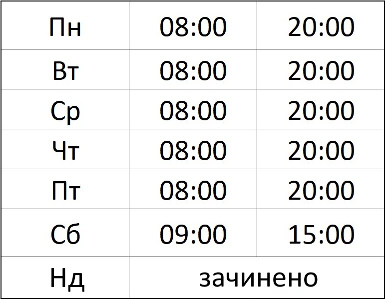
Symptoms and Possible Pathology
Itching and Pain in the Anus
hemorrhoids, fissure, papillitis, periproctitis, fungal and viral lesions (candidiasis, condylomatosis), rarely – a symptom of coccygodynia
Bleeding
hemorrhoids, fissure, polyps and tumors of the intestine, ulcers and diverticula of the intestine
Digestive Disorder, Diarrhea
pancreatitis, individual food intolerance, polyps and tumors of the intestine and abdominal cavity, ulcerative colitis, Crohn’s disease
Constipation, Excretory Dysfunction
Abdominal Pain, Vomiting, Fever
Book a consultation with Oleksandr Kosenko
Modern Diagnosis
Proctological Examination
- General surgical examination (examination of the whole body and mucous membranes), conversation with the patient regarding complaints.
- Digital examination of the anal canal and rectum, with possible examination of the prostate gland in men, examination of the mammary glands, and transvaginal examination in women.
- Anoscopy and rectomanoscopy — examination of the anal canal and rectum using proctological instruments to a maximum depth of up to 30 cm.
Endoscopic Examinations
- EGD (esophagogastroduodenoscopy) — examination of the esophagus, stomach, and duodenum using a flexible video endoscope.
- Colonoscopy — examination of the entire length of the colon using a flexible video endoscope.
- Rectomanoscopy also belongs to the category of endoscopic examinations and allows examination of the rectum to a depth of up to 30 cm. All examinations are performed by physicians of the highest category from the Left Bank Proctology Center.
Radiological Examinations
- Irrigography (proctography) — X‑ray images of various segments of the colon after an enema with a harmless radiopaque contrast agent — barium sulfate.
- Barium passage through the gastrointestinal tract (X‑ray monitoring of the duration of food passage after ingestion of contrast).
- Fistulography — staining of fistulous tracts and cavities with a sterile radiopaque contrast agent with fixation of the image. All X‑ray examinations are performed in the presence of a physician who views the image in real time on the screen and adjusts the X‑ray procedure.
No Answer?
Ask Doctor Kosenko!
Surgeon–proctologist of the highest category Oleksandr Petrovych Kosenko – phone/Viber: +380 50 330 15 15, e‑mail: info@kosenko.org
Diagnosis and treatment of proctological diseases, as well as diseases of general surgery, oncology, gastroenterology, and urogynecology. Performs all types of proctological surgical and minimally invasive procedures, as well as a range of general‑surgical operations. Work experience: 2000–2025. Worked as a surgeon–proctologist at the Ukrainian Center for Coloproctology (metro station “Universytet”, 17 T. Shevchenka Blvd., City Clinical Hospital No. 18), simultaneously as a surgeon in the general surgery department of City Clinical Hospital No. 18, as a surgeon in the purulent surgery department of City Clinical Hospital No. 3 / Sepsis Center, and as a surgeon–proctologist in several private medical centers.
Patients are seen at the following address: metro station “Livoberezhna”, Rusanyvka residential area, 49 Entuziastiv St., “Rusanyvka” polyclinic, on the premises of the medical center “MSC” (Medical Service Consulting). Report to room №1 on the first floor – the first door to the right after entering the polyclinic, or to the reception room – after entering, turn right, the next door with the sign “MEDICAL CENTER MSC” located after room №3. Appointments are conducted by prior booking at a scheduled time arranged personally with the doctor, or via the administrator Tetiana at phone/Viber/Telegram/WhatsApp +38098 955 34 10, or via the senior nurse Liliya at phone/Viber/Telegram +38093 376 70 56.
See prices for a proctologist consultation and proctological examination programs. Home visits to patients are possible within Kyiv and its surroundings (left / right bank / +20 km beyond the city) – 2500–3500 UAH / 50 minutes. Appointments on Saturday: +50% of the cost. Appointments outside working hours or on Sundays: +100% of the cost. Questions regarding types and cost of surgical procedures will be addressed after a detailed examination or review of previous investigations.
To book a consultation, send us a message via Viber (or WhatsApp / Telegram / a regular SMS to the phone number +38098 955 34 10): full name, age, preferred date and time, a brief description of the problem. You may also use the online proctologist appointment booking system. The administrator will advise you on how to prepare properly for the examination.
Phone: +38050 330 15 15, e‑mail: info@kosenko.org
- Attention: we operate even without electricity supply; the operating room is serviced by our own inverter batteries.
- Download the patient preparation instructions for a proctologist examination.
Certificates of Doctor Kosenko Oleksandr Petrovych
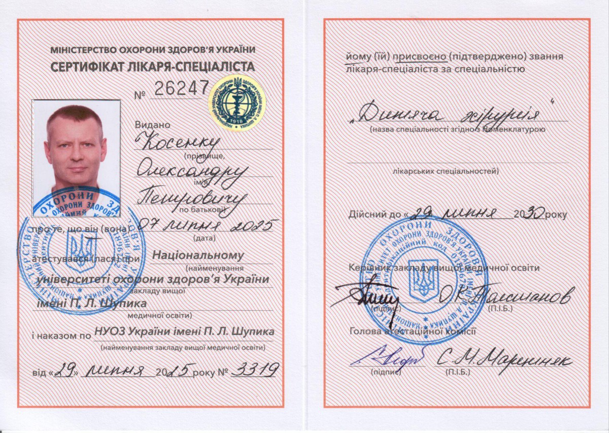
Сертифікат Олександра Косенка. Сертифікат лікаря-спеціаліста з дитячої хірургії
Сертифікат Олександра Косенка. 12 друзів андрології. 2025 рік
Сертифікат Олександра Косенка. Актуальні питання невідкладної хірургії. 2025 рік
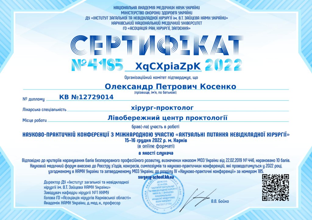
Сертифікат Олександра Косенка. Актуальні питання невідкладної хірургії. 2022 рік
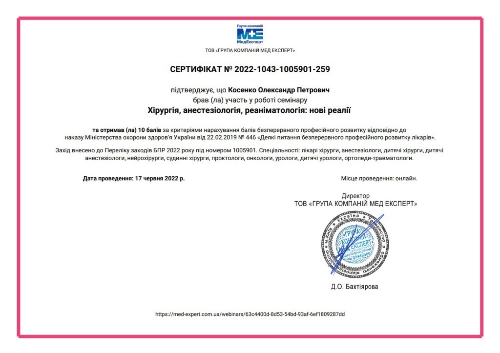
Сертифікат Олександра Косенка. Хірургія, анестезіологія, реаніматологія: нові реалії. 2022 рік
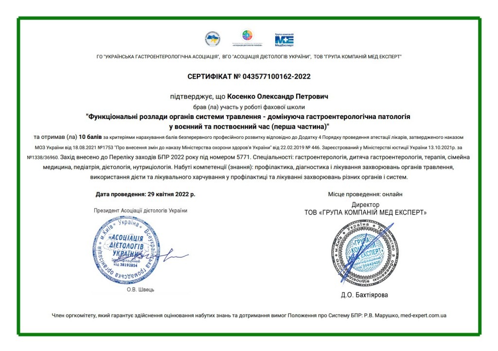
Сертифікат Олександра Косенка. Функціональні розлади органів системи травлення – домінуюча гастроентерологічна патологія у воєнний та поствоєнний час (перша частина). 2022 рік
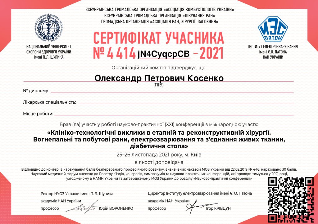
Сертифікат Олександра Косенка. Клініко-технологічні виклики в етапній та реконструктивній хірургії. Вогнепальні та побутові рани, електрозварювання та з’єднання живих тканин, діабетична стопа. 2021 рік
Сертифікат Олександра Косенка. Менеджмент больового синдрому. 2021 рік
Сертифікат Олександра Косенка. International Summit on Anal Fistula. 2021 рік
Сертифікат Олександра Косенка. Актуальні питання невідкладної хірургії. 2021 рік
Сертифікат Олександра Косенка. Клініко-технологічні виклики в етапній та реконструктивній хірургії. Вогнепальні та побутові рани, електрозварювання та з’єднання живих тканин, діабетична стопа (в онлайн режимі). 2020 рік
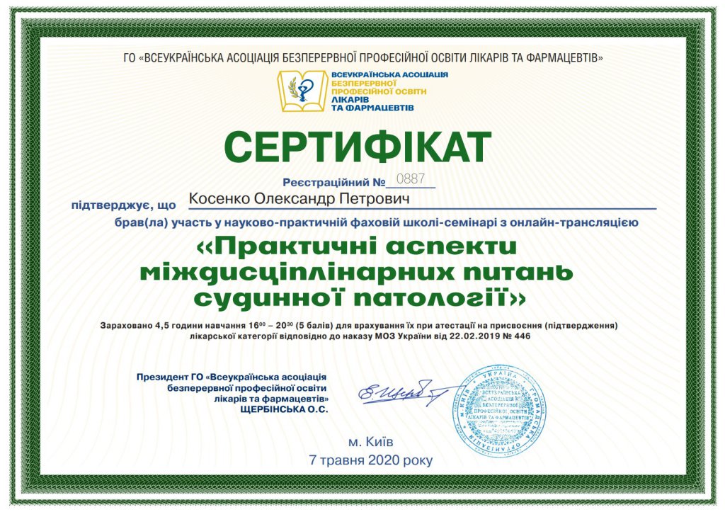
Сертифікат Олександра Косенка. Практичні аспекти міждисциплінарних питань судинної патології. 2020 рік
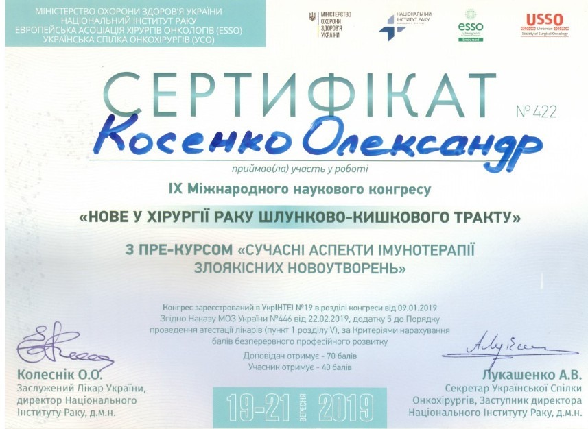
Сертифікат Олександра Косенка. Нове у хірургії раку шлунково-кишкового тракту. 2019 рік
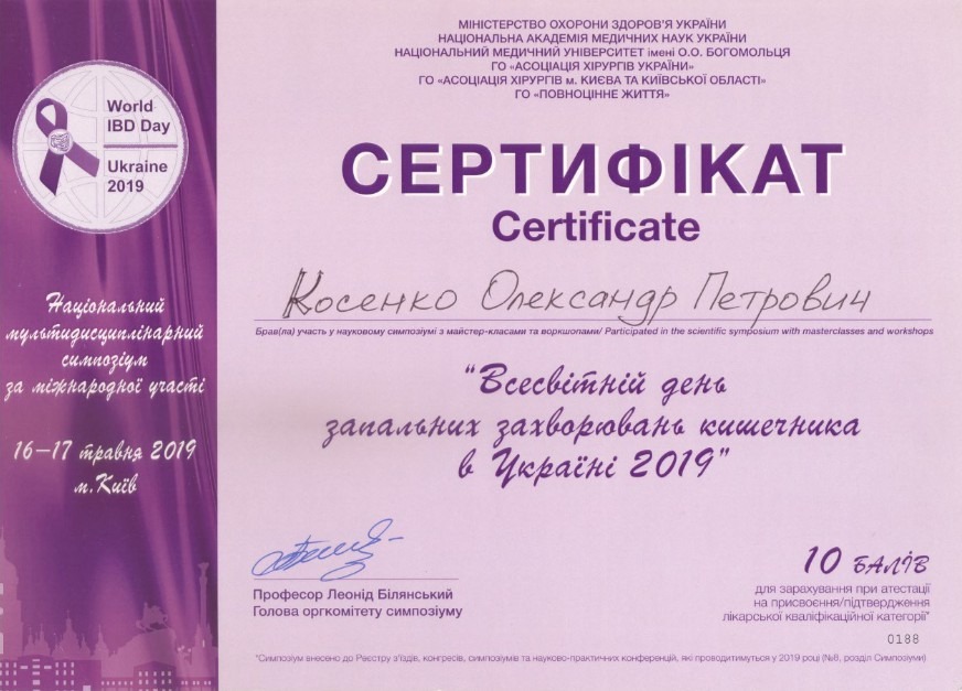
Сертифікат Олександра Косенка. Всесвітній день запальних захворювань кишечника в Україні. 2019 рік
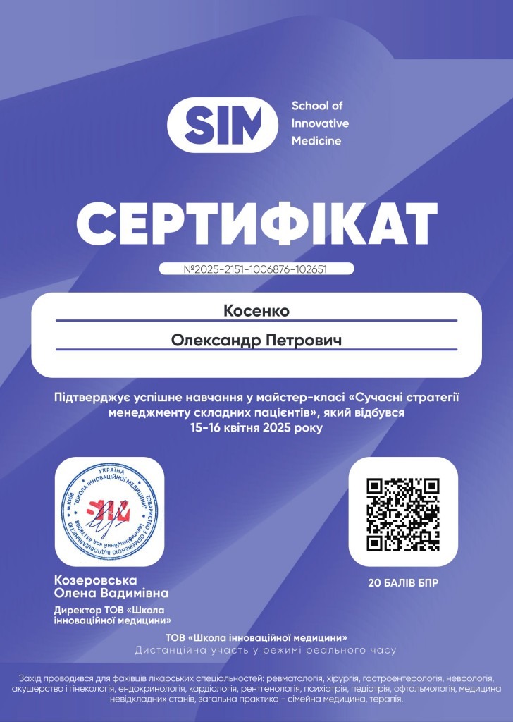
Сертифікат Олександра Косенка. Сучасні стратегії менеджменту складних пацієнтів. 2025 рік
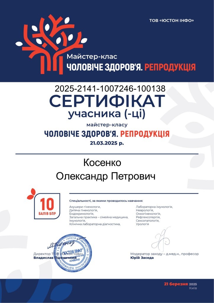
Сертифікат Олександра Косенка. Чоловіче здоров’я. Репродукція. 2025 рік
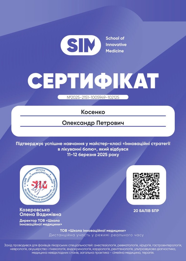
Сертифікат Олександра Косенка. Інноваційні стратегії в лікуванні болю. 2025 рік
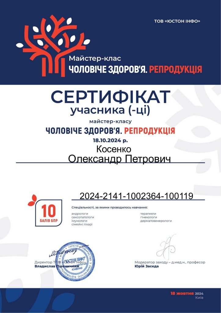
Сертифікат Олександра Косенка. Чоловіче здоров’я. Репродукція. 2024 рік
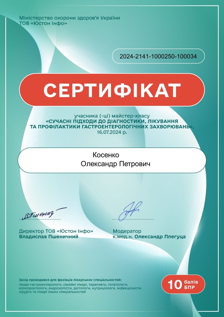
Сертифікат Олександра Косенка. Сучасні підходи до діагностики, лікування та профілактики гастроентерологічних захворювань. 2024 рік
Сертифікат Олександра Косенка. Shifting the paradigm of providing comprehensive medical care in wartame in conditions of limited resources. CNS disorders during wartime (traumas, concussion, chemical poisoning). 2022 рік
Сертифікат Олександра Косенка. Школа сучасної урології та андрології. 2022 рік
Подяка Олександру Косенку. За високий рівень професіоналізму, вагомий внесок в охорону здоров’я громадян вільної України та з нагоди Дня медичного працівника. 2018 рік

Сертифікат Олександра Косенка. Інновації в хірургії ран, з’єднань. Застосування електрозварювання живих тканин. 2014 рік

Сертифікат Олександра Косенка. Інновації у хірургії ран, з’єднань та стопи діабетика. 2014 рік

Сертифікат Олександра Косенка. Хірургія поранень. Застосування електрозварювання тканин. 2014 рік
Сертифікат Олександра Косенка. Курс проблем державного управління. 2014 рік

Сертифікат Олександра Косенка. Перспективи лікування пухлин шлунково-кишкового тракту. 2012 рік

Сертифікат Олександра Косенка. 6-й міжнародний курс ендоскопічної хірургії. 2015 рік

Сертифікат Олександра Косенка. Мінімально-інвазивна хірургія органів малого тазу. 2017 рік

Сертифікат Олександра Косенка. Курс трансанальної гемороїдальної деартеріалізації. 2011 рік

Сертифікат Олександра Косенка. Лапароскопічні технології сьогодні. 2010 рік

Сертифікат Олександра Косенка. Сучасні технології в хірургії раку шлунково-кишкового тракту. 2011 рік
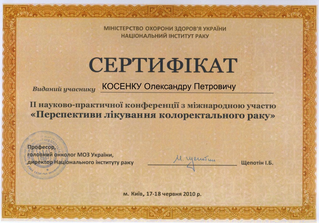
Сертифікат Олександра Косенка. Перспективи лікування колоректального раку. 2010 рік

Сертифікат Олександра Косенка. Лапароскопічні технології у сучасній хірургії. 2009 рік

Сертифікат Олександра Косенка. Перспективи лікування колоректальних тканин. 2008 рік

Сертифікат Олександра Косенка. Рани м’яких тканин. 2008 рік

Сертифікат Олександра Косенка. 2-й з’їзд колопроктологів. 2006 рік
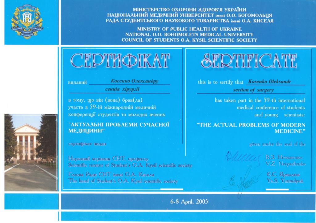
Сертифікат Олександра Косенка. Актуальні проблеми сучасної медицини. 2005 рік
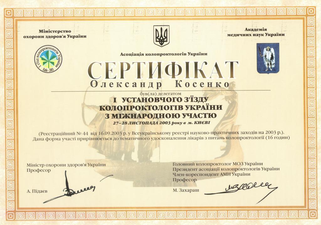
Сертифікат Олександра Косенка. 1-й з’їзд колопроктологів. 2003 рік
Legal Details and Information
| Taxpayer Identification Card Number | 2844108636 |
| Location of the Sole Proprietor | Ukraine, 02002, Kyiv, Raіsa Okipna St., building 3-V, apartment 18 |
| Date and Number of Entry in the Unified State Register of Legal Entities, Individual Entrepreneurs and Public Formations | 23.01.2024, 2000670010001180188 |
| Types of Economic Activity | 68.20 Provision of Rental and Operation of Own or Leased Real Estate (primary) 77.39 Rental of Other Machinery, Equipment and Goods n.e.c. 96.09 Provision of Other Personal Services n.e.c. 96.04 Physical Well-Being Services 86.90 Other Human Health Activities 86.22 Specialized Medical Practice Activities 86.21 General Medical Practice Activities 82.99 Other Business Support Service Activities n.e.c. 77.40 Leasing of Intellectual Property and Similar Products, Except Copyrighted Works |
| Download extract of Sole Proprietor Kosenko O.P. | |
| Bank Details | |
| Bank Name | Raiffeisen Bank JSC |
| MFO | 380805 |
| IBAN | UA803003350000000026005699674 |
| Account Currency | UAH (hryvnia) |
| Account Opening Date | 04.12.2024 |
Use of Terms and Privacy Policy for the kosenko.org Website
Doctor: Kosenko Oleksandr Petrovych
1.1. About this Document
This document contains the Terms of Use and Privacy Policy (hereinafter – “Document”) that regulate the use of the website kosenko.org (hereinafter – “Site”), owned by Doctor Kosenko Oleksandr Petrovych (hereinafter – “Doctor”, “we”). Use of the Site means full agreement with the terms of this Document.
1.2. Definition of Terms
-
“User” – a natural person who uses the Site, receives information or medical services through it.
-
“Personal Data” – information or a set of information about a natural person who is identified or can be specifically identified.
-
“Medical Information” – information about health status, diagnoses, examination results, treatment, medical history.
-
“Medical Confidentiality” – confidential patient information that is subject to protection in accordance with Ukrainian legislation.
-
“Processing of Personal Data” – any action or set of actions with personal data.
-
“Cookies” – small text files stored on the user’s device when visiting the site.
1.3. Scope of Application
The Document applies to all interactions with the Site, including viewing information, using online services, obtaining consultations and other medical services.
2.1. Rights and Obligations of the User
The User has the right:
-
to receive reliable information about medical services;
-
to demand compliance with medical confidentiality;
-
to control the processing of their personal data;
-
to seek consultations within the limits established by law.
The User undertakes:
-
to provide reliable information about their health status;
-
to comply with the rules of using the Site;
-
not to use the Site for unlawful purposes;
-
to respect the rights of other users and medical personnel.
2.2. Usage Restrictions
The following is prohibited:
-
distributing spam or fictitious information;
-
posting content of a sexual nature or with obscene expressions;
-
posting threatening or offensive content;
-
violating copyrights and intellectual property rights;
-
impersonating another person;
-
using the Site for advertising purposes without permission;
-
attempting to gain unauthorized access to the system.
2.3. Online Medical Consultations
Medical online informing is carried out in accordance with Article 78 of the Basic Legislation of Ukraine on Health Care as the dissemination of scientific and medical knowledge among the population. Online consultations do not replace a full medical examination and treatment.
2.4. Disclaimer
The Site and the information posted on it are provided “as is” without any warranties. The Doctor is not responsible for:
-
technical failures in the operation of the Site;
-
inability to use certain functions;
-
actions of third parties;
-
consequences of self-treatment if the user uses information from the Site without a personal consultation with the doctor.
3.1. Purpose of Processing Personal Data
Personal data are processed for the following purposes:
-
identification of users and provision of medical services;
-
maintenance of medical documentation in accordance with legal requirements;
-
ensuring security and preventing fraud;
-
improving the quality of medical services;
-
fulfilling legal obligations.
3.2. Types of Personal Data that May Be Processed:
-
surname, first name, patronymic;
-
date of birth;
-
taxpayer identification card number (if available);
-
phone number and email;
-
residential address;
-
identity documents;
-
data of the legal representative (for children or persons who cannot act independently);
-
information about health status, medical history, examination results, treatment results, prescriptions;
-
history of appeals, consultations, referrals, extracts, electronic prescriptions, medical reports, immunizations, hospitalizations, procedures, treatment plans;
-
medical images, videos, diagnostic test results;
-
insurance information (if available);
-
consent to the processing of personal data, selected authentication method;
-
IP address, browser information, operating system, time and duration of visit, viewed pages, referring site (site-referrer), search queries, other technical parameters used for traffic analysis and improving the Site’s operation;
-
other personal data directly provided for by law or necessary for providing medical services and maintaining medical documentation.
3.3. Legal Basis for Processing
Processing of personal data is carried out on the basis of:
-
user consent;
-
necessity to fulfill the contract for the provision of medical services;
-
fulfillment of legal obligations;
-
protection of vital interests of the user or another person.
3.4. Protection of Medical Confidentiality
The Doctor undertakes to comply with medical confidentiality in accordance with Ukrainian legislation. Medical information may be disclosed only:
-
with the patient’s consent;
-
in cases established by law;
-
by court decision;
-
in the interests of national security.
3.5. Use of Cookies
The Site uses necessary, functional, and analytical cookies to ensure operation, improve usability, and analyze traffic. You can manage cookies through browser settings. Disabling cookies may affect the Site’s functionality.
3.6. Transfer of Data to Third Parties
Personal data may be transferred to:
-
other medical specialists for consultations;
-
laboratories for conducting tests;
-
authorized state authorities in cases established by law;
-
with your written consent – to other persons.
3.7. International Data Transfer
In case of using international services or transferring data outside Ukraine, an appropriate level of personal data protection is ensured in accordance with legal requirements.
3.8. Data Storage Periods
-
Medical records of inpatients are stored for 25 years.
-
Medical records of outpatients are stored for 5 years.
-
Other types of documentation are stored in accordance with current legislation and the List of Standard Documents.
4. RIGHTS OF PERSONAL DATA SUBJECTS
4.1. Your Rights
You have the right:
-
to receive information about the processing of your personal data;
-
to receive information about the processing of your personal data free of charge;
-
to demand changes to inaccurate or incomplete data;
-
to demand deletion of personal data in cases provided by law;
-
to demand suspension of processing in certain cases;
-
to object to data processing on certain grounds.
4.2. How to Exercise Your Rights
To exercise your rights, contact:
-
by phone: +38050 330 15 15;
-
by email: info@kosenko.org;
-
in writing to the address: 02147, Kyiv, Entuziastiv St., 49, office 3, to Kosenko Oleksandr Petrovych;
-
during personal reception.
The request must contain your identification data and a description of the requested information or action.
5.1. Technical and Organizational Measures
-
data encryption during transmission and storage;
-
regular backup;
-
antivirus protection;
-
access control to servers;
-
appointment of a person responsible for personal data protection;
-
staff training on data protection issues;
-
regular security measures audit.
5.2. Security Incidents
In case of personal data security breach:
-
immediate measures are taken to stop the breach;
-
possible consequences are assessed;
-
relevant authorities are notified within the established timeframes;
-
affected persons are informed.
6. ADDITIONAL REQUIREMENTS UNDER UKRAINIAN LAW
6.1. Notification to the Authorized Person on Human Rights
In accordance with Art. 9 of the Law of Ukraine “On Personal Data Protection”, the doctor notifies the Authorized Verkhovna Rada of Ukraine on Human Rights about the processing of personal data that pose a special risk to the rights and freedoms of data subjects within 30 working days from the start of such processing.
6.2. Person Responsible for Personal Data Protection
The person responsible for organizing work on personal data protection is Kosenko Oleksandr Petrovych. Contact details: +38050 330 15 15, info@kosenko.org.
6.3. Compliance with Medical Advertising Requirements
The Site contains both informational and educational materials and information about medical services. All medical services advertising is placed in accordance with the requirements of the Law of Ukraine “On Advertising” and does not contain guarantees regarding treatment results.
6.4. Electronic Healthcare System (EHS)
The site kosenko.org and Doctor Kosenko Oleksandr Petrovych are connected to EHS in accordance with current legislation requirements.
6.5. Criminal Liability
Disclosure of medical confidentiality entails criminal liability under Art. 145 of the Criminal Code of Ukraine.
7.1. Pre-trial Settlement
Before applying to court, it is mandatory to submit a claim in writing or electronically. Claim consideration – up to 30 calendar days.
7.2. Judicial Dispute Resolution
If pre-trial settlement is impossible, the dispute is considered in the courts of Ukraine.
7.3. Applicable Law
Ukrainian legislation applies to relations between the parties.
8. INFORMATION ABOUT DOCTOR AND SITE
-
Doctor: Kosenko Oleksandr Petrovych
-
Address: 02147, Kyiv, Entuziastiv St., 49, office 3
-
Phone: +38050 330 15 15
-
Email: info@kosenko.org
The Doctor reserves the right to amend this Document. The new version takes effect from the moment of its publication on the Site. Users are notified of significant changes through the Site or email (with consent).
Last Update: 2025.09.12
Publication Date: 2025.09.12